
Microscopes and digital technology in dentistry
Originally published
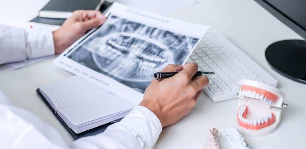
Dental technology can help a clinician see detail, record the shape of teeth and plan treatment. Different tools answer different questions: a microscope enlarges what can be seen, an optical scanner records tooth surfaces, and an X-ray shows structures that are not visible directly.
The useful question is what a particular device adds to your care. Equipment supports clinical judgement; it does not guarantee a painless procedure, a perfect fit or a lasting result.
Seeing fine detail with a microscope
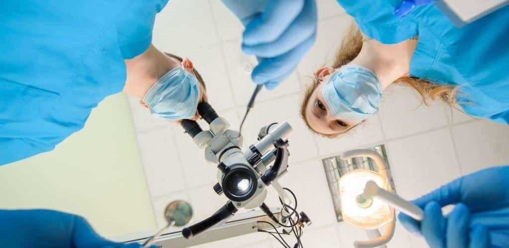
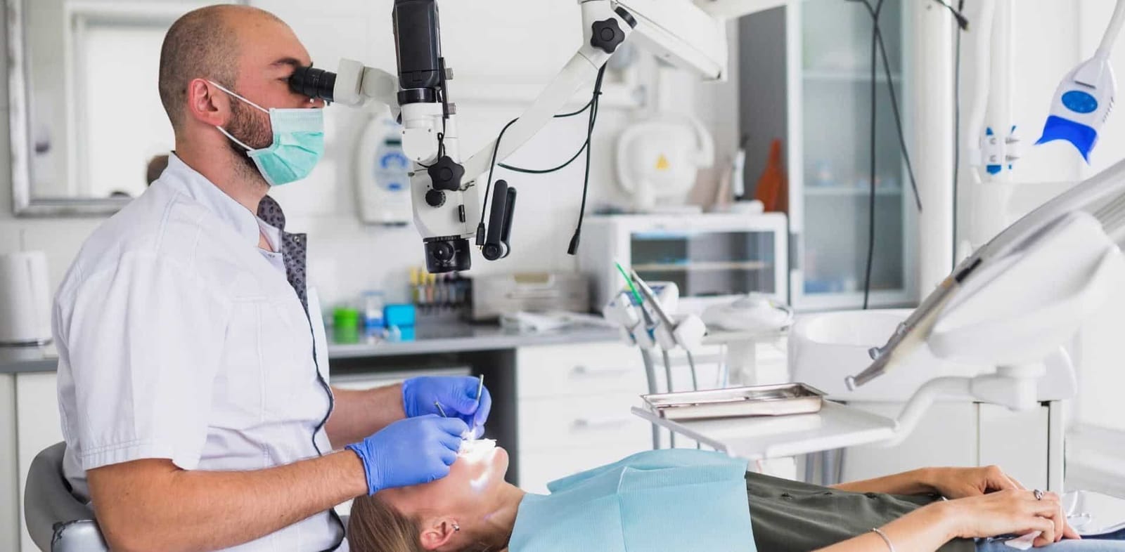
A dental microscope combines magnification with lighting. It can help a clinician inspect small structures that are difficult to see directly. The magnification used varies with the equipment and the task, rather than always being ten times normal size.
Our practice uses a microscope in dental treatment. Your dentist can explain whether it is useful for the procedure being considered.
How does it help treatment?
Microscopes are particularly useful in root canal treatment. They can help locate small canal openings, assess fine details and manage some problems during retreatment. Better visibility can help the clinician preserve tooth tissue, but it does not make every canal accessible or every tooth treatable.
Diagnosis still combines your symptoms, examination, tests and appropriate imaging. A microscope cannot detect every early problem or replace an X-ray where one is needed. Treatment time, cost and outcome depend on much more than magnification.
Magnification can also help when checking accessible tooth surfaces and restoration margins. A filling’s condition, the remaining tooth, your bite and ongoing care all affect its performance. No microscope makes a filling immune to decay or guarantees that it will never need replacement.
It is also misleading to promise that technology leaves no bone fragments after extraction, speeds every wound’s healing or ensures perfect implant positioning. Surgical planning, technique, anatomy and recovery all matter.
Digital impressions and treatment planning
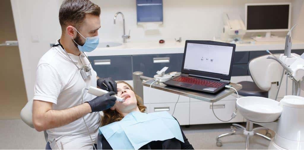
An intraoral scanner records the visible surfaces of teeth and gums to build a digital model. This can be used in planning or making some crowns, bridges and orthodontic appliances. It is different from a CT scan and does not use X-rays.
Some patients prefer scanning to an impression tray, particularly if impression material makes them gag. Conventional impressions remain useful, and neither method is automatically inaccurate or uncomfortable. The best choice depends on the work required and the quality of the record.
A digital model or simulation can help you discuss proposed changes. It is a planning aid, not a promise that your teeth will look exactly like the image. Ask what is predictable, what may change and whether the plan involves removing healthy tooth tissue.
X-rays, cameras and lasers
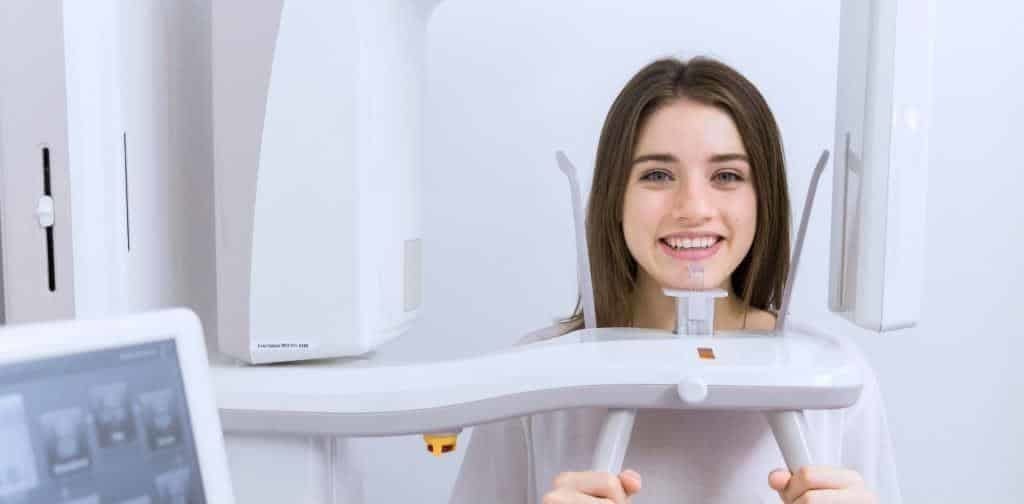
Cone beam CT, or CBCT, produces three-dimensional X-ray images. It may help when ordinary dental X-rays cannot answer a particular question, such as the relationship between a tooth and nearby structures. It does not routinely replace standard dental X-rays.
CBCT uses ionising radiation. The dose depends on the equipment, settings and area scanned, and is generally higher than for conventional dental radiographs. It should therefore be justified by the information needed, rather than described simply as a minimal-dose upgrade for everyone.
An intraoral camera records photographs inside the mouth and can help your dentist explain a finding. Dental lasers have selected uses that depend on the device and the procedure; they are not a universal replacement for other instruments or anaesthesia. These examples do not mean that every device or procedure is available at this practice.
Ask our team how the proposed equipment will help with your treatment, what alternatives exist and how the result will be checked.